Biological Observations
Almost half of the scientific papers of Giovanni Battista Amici were written about biological observations, mostly about botany (morphology, histology, physiology, fertilization, embryology and phytopathology) but also about animal histology (muscular fibre).
In more than forty years of microscopic observations in the field of botany his interests were concentrated on four main issues: circulation of sap in the Chara cells; morphology of the Characeae; fertilization process in the Phanerogamous plants (Angiosperms); phytopathologies.
Circulation of the sap and morphology of the Charæ
In the opening lines of the paper of 1818 Osservazioni sulla circolazione del succhio nella Chara < Observations on the Circulation of Sap in the Chara > (“Memorie di Matematica e di Fisica della Società Italiana delle Scienze”, vol. XVIII-1820, download pdf ), Amici immediately declared that his biological investigations were dependent on the construction of his catadioptric microscope:
As soon as I had a Catadioptric Microscope which I had made for myself, I wished to collect objects which for their small size permitted judging the force of the instrument better, or that could satisfy my curiosity due to their singular structure, and which at the same time promised some useful discovery in their organization.
Among the many I chose my attention was mainly attracted by the Chara vulgaris, a small aquatic plant in which our Abbé Corti had discovered a circulation of sap as early as 1774. This particular phenomenon of the visible rising and descending of the fluid struck me so strongly that I decided to institute a series of experiments on this plant.
The main questions clarified by this first research were
the dependence of this circulation on the series of grains of chlorophyll, the greater rapidity of flow of the sap near the cell walls than in the depth of the cell, the lack of a dividing wall between the rising and descending flow, the possibility of dividing the current of the sap in two separate circuits by constricting the cell (H. von Mohl, Giambattista Amici, 1863).
Amici continued to study the Charæ for a long time, as can be seen from his papers:
Osservazioni microscopiche sopra varie piante (1822), Descrizione di alcune specie nuove di Chara ed osservazioni microscopiche sulle medesime (1827), Sulla circolazione che si osserva negli internodi della Chara (1839), Osservazioni sugli zoospermi della Chara (1842) < Microscopic observations on various plants, Description of some new Chara species and microscopic observations of the same, On the circulation that can be seen in the internodes of the Chara, Observations on the zoosperms of the Chara >.
In the paper of 1822 he wrote about his research in vegetable histology as well
(cf. Microscopic observations on various plants, the fourth article, Dell’Epidermide < Of the Epidermis >, and the fifth article, Dell’unione del tessuto vegetale
< Of the union of vegetable tissue >).
Discovery of the pollen tube
The investigations of historic importance of Giovanni Battista Amici regard the process of fertilization. The first observation (1822), which led to the fundamental discovery of the pollen tube, was by chance (Microscopic observations on various plants, third article, On Pollen, download pdf ). While investigating the hairs on the stigma of the Portulaca oleracea to see if it wasn’t possible to find sap circulation like that present in the cells of the Chara - as was indeed the case - I happened, wrote Amici,
to observe a hair with a grain of pollen attached to its tip which after some time suddenly exploded and sent out a type of transparent gut which stretched out along the hair and joined it laterally.
Studying this new organ with attention, I realized that it was a simple tube composed of a subtle membrane, so I was quite surprised to see it full of small bodies, part of which came out of the grain of pollen and the others which entered after having travelled along the tube or gut.
This discovery was the first step towards the correct explanation of the process of fertilization. The second, mentioned in a letter to Charles-François Brisseau Mirbel (1776-1854) in July 1830, was based on observations made on the Hibiscus syriacus and on the pumpkin (Note sur le mode d’action du pollen sur le stigmate, downloadl pdf ): “C’est le boyau lui-même qui peu à peu s’allonge, descende par le style, et va se mettre en contact avec l’amande”.
Amici was becoming convinced that “the pollen gut which has penetrated the conducting tissue continues to lengthen out until it is inside the ovary, where it meets with the esostoma of the ovules, without breaking into the conducting tissue, and which was a new proof of the conservation of the gut in its integrity” (Sul processo col quale gli ovuli vegetabili ricevono l’azione fecondante del polline < On the process by which the vegetable ovules receive the fertilizing action of the pollen >, 1839).
 Eight years passed between the first discovery and the letter to Mirbel about this phenomenon. In the meantime, Adolphe Brongniart (1801-1876) had found that “the exit of tubes from the grains of pollen is a completely generalized phenomenon and that the pollen guts penetrate in the conducting tissue of the stigma and of the style”. In 1831-1833 Robert Brown (1773-1858) published his research on the fertilization of Orchids and Asclepiads, but it did not also describe the transformations which take place in the ovule, and therefore remained behind when compared to Amici’s research.
Schleiden, for his part, with great energy and with a great number of investigations about the structure of the ovule and the transformations which take place in it, had tried to determine how the embryo was first formed, but he had taken the wrong path and this produced a great deal of confusion in the world of botany (H. von Mohl, Giambattista Amici, cit.).
Fertilization of plants: Cucurbita Pepo
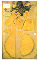 At the First Meeting of Italian Scientists in Pisa in 1839 he seems to have had the crowning touch of an important phase of his investigations with the presentation of a wax model, created in the laboratories of the Royal Museum by Luigi Calamai (1800-1851), reproducing the phenomenon as he believed he had seen it in the Cucurbita Pepo (Sul processo col quale gli ovuli vegetabili ricevono l’azione fecondante del polline).
At the end of 1839 Amici was still unprepared to face the issue, but three years later he felt ready to offer valid observations in opposition to the theory of Matthias Jacob Schleiden (1804-1881).
In the Fourth Meeting of Padua on 1842 he believed he was able to offer a complete solution to the problem “since he had managed to point out that in the pumpkin (Cucurbita pepo) the embryo develops from a body pre-existing in the ovule, and this body absorbs the fertilizing secretion which the gut transmits to it” (Sulla fecondazione delle piante <Cucurbita pepo> (On the fertilization of plants <Cucurbita pepo>]). In reality Cucurbita was not very appropriate for the demonstration Amici intended to make, because it behaves anomalously even in comparison to other cucurbits. For this reason the proofs offered against the theories of Schleiden were not in reality very convincing. The demonstration would follow, full and faultless, only four years later, when Amici would base it on the Orchids. He was in any event the first who was able to make others listen, affirming that the pollen gut did not transform itself into a germinating vessel. But his presentation in 1842 provoked
an attack from Schleiden (Flora 1845) in most violent terms. This response left Amici perfectly calm; he wrote me that he knew he had seen correctly, that surely sooner or later someone would have confirmed his observations, that he was a peaceful person and so he would not respond to Schleiden. In recompense, he received a splendid satisfaction. A few months later he produced a microscope for Schleiden which would have allowed this latter able to observe better, and Amici demonstrated the correctness of his own theories on the fertilization through new research made on Orchids
(H. von Mohl, Giambattista Amici, cit.).
Fertilization of Orchids
Does fertilization in phanerogamous plants take place in the way that Schleiden claims, that is, with the point of the pollen gut which penetrates the integument of the ovule, and pushing back the membrane of the embryonic sac forms a cavity to remain there and subsequently change into the real embryo?
The special research which I carried out on the pumpkin (cucurbita pepo) convinced me that in this plant the fertilization takes place in a completely different way.
The third and final step towards the correct explanation of the process of fertilization is constituted by a paper of historical importance, Sulla fecondazione delle orchidee <On the fertilization of Orchids> (download pdf ), which Amici presented at the Eighth Meeting in Genoa in 1846; he had the conclusions of this paper represented some time later by a wax model created with every probability by the same Calamai.
For the first time in this work by Amici a correct and coherent exposition of the overall process of fertilization of a plant in all its stages was given, from the pollination of the stigma up to the full development of the embryo. It was a model for all subsequent research and at the same time the final blow to Schleiden’s mistaken
theory [...]
Amici [...] also followed the further processes in the ovule of the Orchids, the existence of the germinative vesicle before the fertilization, the formation and the development of the embryo from this, in such a complete way that subsequent research by others could only furnish a confirmation but not important addition (H. von Mohl, Giambattista Amici, cit.).
Phytopathology
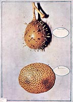
When the grapevine cryptogam from the farms of Margate in England reached Tuscany at the beginning of the 1850s, Giovanni Battista Amici and Filippo Parlatore (1816-1877) were included in the Commission formed to study the causes of the scourge. Amici
provided a good description of the so-called Oidium Tuckeri and so added to the progress in the knowledge of the organization of the Erysiphe, by discovering the way of fructification - until then unknown - as much in grape fungi as in other Erysiphe species, which gave Ehrenberg the opportunity to enunciate the Cicinobolus genus. But he did not see the relation of this fructification to those already known in Erysiphe, and for this reason he explained that the Oidium Tuckeri could yes, possibly but not probably, be an Erysiphe. Regarding the question which was hotly debated at that time in Italy, whether the fungus was the cause or the effect of the illness, he was on the side of those who supported this latter opinion (H. von Mohl, Giambattista Amici, cit.).
 Amici presented many papers and announcements on this subject to the Academy of the Georgofili in Florence: Osservazioni microscopiche sulla malattia dell’uva
< Microscopic observations on the disease of the vine > (1852), Sulla malattia dell’uva, Memoria < On the disease of the vine >, Paper (1852) (download pdf), Lettera sull’Oidium Tuckeri < Letter on the Oidium Tuckeri > (1853), Lettera intorno all’oidio della vite
< Letter about the oidium in grapevines > (1854), Lettera intorno alla crittogama della vite < Letter about the grapevines cryptogram > (1856).
In the field of phytopathology he also investigated rachis in wheat, the withering of the mulberry leaf, the lime disease of silkworms.
Animal Histology
In 1858 Amici published the paper Sulla fibra muscolare < On muscle fibre > (download pdf) in the Journal for Medicine, Surgery and Related Sciences “Il Tempo” of Florence; it is interesting for a number of reasons.
For thirty years now - he wrote - I have looked for natural objects whose delicate structures could be used as paragons for the optical power of my microscopes. It was then that among the many test objects that I noted, fly muscle fibre seemed to me to possess particularities fine enough to fully satisfy the purpose I had set for myself [...]
It was already known that voluntary muscle fibre from animals contains equidistance, transversal lines, or striae, more or less close to each other depending on muscle tension, and more or less visible depending on the animal. No one, however, had discovered the existence of the very subtle threads which unite one stria with another longitudinally, as I saw there was in the fibre from the fly. The observation was rather difficult and I believe that in that period demonstrating the presence of the threads with certainty was a property of very few microscopes, perhaps only of the one that Lister possessed.
Amici charged Egisto Tortori (1829-1893) - a student of Calamai and the last representative of the Florence school of wax modellers which had developed around the Royal Museum of Physics and Natural History - to model the muscular fibre “showing him, with the help of the microscope, everything I knew about the structure of the voluntary fibre of different animals”.
 In his Nota to this Paper, in 1964, Paolo Buffa spoke of an exceptional correctness in the conclusions of Amici about the structure of the striated muscle fibre. Correctness which was surely due on one hand to the long experience of the observer, and on the other to the superiority of the optical instrument utilized, which from a handwritten note seems to have been an oil-immersion objective. Comparing the images published by John Quekett (A practical treatise on the use of the microscope, Bailliere, London 1848), and by Albert Kölliker (Éléments d’histologie humaine, Masson, Paris 1856) with those of the lamb fibril published by Amici, Buffa declared that
In the light of our current knowledge the superiority of Amici’s images are immediately evident. The illustrations by Kölliker (Fig. 8-A, B and C) show bands of fibril and isolated fibril of muscles from various animals with equidistant transversal striae of equal intensity and thickness; the bands A and I and the line Z are completely missing. The image published by Quekett, in 1848 (Fig. 2 and 8-D), is already much richer in particulars and closer to reality. The fibrils have a structure with alternate light and dark bands (bands I and A) and in one fibril (indicated in
the illustration) it can be seen that the light band is divided in two by a thin dark stria, line Z. It should be noted that the illustrations of Kölliker are enlarged 350 and 600 times, Quekett’s 1200 times and Amici’s 2000 times.
But in his opinion
Amici’s Paper stands out not so much for the sole description of line Z, without doubt more accurate and correct than Quekett’s, as for the whole of original observations on the constitution and structure of the striated muscle fibre and, particularly, for the description of parallel longitudinal threads in the fibril.
Bibliography
Hugo von Mohl, Giambattista Amici, “Botanische Zeitung” (Leipzig), 21. Jahrgang, N. 51, 18. December 1863, p. 1-8; Italian translation by A. Meschiari, “Atti della Fondazione Giorgio Ronchi”, 2-1999; Giovanni Briosi, Giovanni Battista Amici. Cenno sull’opera sua, e ritratto, “Atti dell’Istituto botanico dell’Università di Pavia”, II Serie, vol. XI-1908; Roberto Savelli, Giovan Battista Amici, botanist, “Physis”, VII-1965, fasc. 1, p. 5-47; Paolo Buffa, Note alla Memoria “Sulla fibra muscolare” di Giovan Battista Amici, CNR, Rome 1964; P. Buffa, On the accuracy of the wax models of biological microscopical preparations of Giovan Battista Amici (1786-1863), in Atti del I Congresso Internazionale sulla Ceroplastica nella Scienza e nell’Arte, Florence 3-7 June 1975, Olschki, Florence 1977, p. 217-244; Julius Sachs, Geschichte der Botanik vom 16. Jahrhundert bis 1860, Oldenbourg, Munich 1875 (reprinted 1965), in particular Drittes Buch, Erstes Kapitel, Geschichte der Sexualtheorie; AA.VV., Plant embryology. The Tuscan contribution, Proceedings of the Conference held in Pisa, Department of Botany, 25th October 1986, in honour of the bicentenary of the birthday of Giovan Battista Amici (1786-1863), edited by Fabio Garbari and Ettore Pacini, Pacini Editore, Pisa 1987; A. Meschiari, Corrispondenza di G. B. Amici con scienziati europei (letters < some only partial > with J. Fraunhofer, O. F. Mossotti, J. F. W. Herschel, J. E. Purkyne, H. Mohl, M. J. Schleiden, W. Hofmeister), “Giornale critico della filosofia italiana”, Sept.-Dec. 1997; National Edition of the Works and Correspondence of Giovanni Battista Amici, ed. by A. Meschiari, vol. I, Edited scientific papers, Bibliopolis, Naples 2006.