Instruments
Achromatic microscope
1824 is universally recognized as the year of the birth of the modern compound achromatic microscope. Giovanni Battista Amici was one of the protagonists on an international level of this event and of the later improvements made to the instrument. The English optician Joseph Jackson Lister wrote to Amici on 28 March, 1828:
It is curious that acromatic glasses of short focus should have been produced (as appears) near the same time in Italy & England, independent of each other. From some circumstances I suspect that those of Chevalier in France were undertaken in consequence of the report of their successful production elsewhere. (I think it was in 1824 that Tulley executed his first good triple 0,4 inch). I should ful obliged to thu to inform me how long they have been employed by thu, whether any one on the continent before thu had succeeded in them, and also (which I think I did hear from thu but I have forgotten it) to whom we are originally indebted for the important improvement of placing one before another in combination. We knew nothing of its value here till I had over from France one of Chevalier’s Instruments, & had enlarged its apertures (cf. A. Meschiari, The microscopes of Giovanni Battista Amici, Tassinari, Firenze 2003).
 See also J. J. Lister, On some properties in achromatic object-glasses applicable to the improvement of the microscope (“Philosophical Transactions of the Royal Society of London” for the year 1830).
Amici wrote back, on 12 May:
As for your request to know the period in which I first worked on achromatic microscope object-glasses, I can tell you that it was towards 1815 when I fitted my large-aperture Newtonian telescopes with achromatic eye-pieces. My greatest success consisted of an object-glass composed of two French glass convex lenses with between them a concave lens made of old English flint-glass having the following radii of curvature expressed in lines of Paris feet, commencing towards the object: 5 : 5 : 5 : 2.5 : 3.5 : 5.5, and focal length more than seven lines, but it was not the equal of my reflecting microscope. Although the illustrious Fraunhoffer had some time previously placed achromatic microscopes on the market, he was no more advanced than me. It was only in the year 1824 that I returned to this work with the certainty of greater success after reading the report made by Mr. Fresnel to the Académie Royale des Sciences in Paris about the achromatic microscope of M. Selligue. It was from this latter that I took the fortunate idea of combining various achromatic object-glasses [read: lenses] instead of constructing just one of focal length equal to that of their combination [...]
 In 1827 during his first trip to Paris and London he brought his new achromatic microscope with him. Shortly after he left England, the Notice of some Microscopic Observations of the Blood and Animal Tissues, by Dr. Hodgkin and J. J. Lister, communicated by Dr. Hodgkin, appeared in “The Philosophical Magazine and Annals of Philosophy” (N° 8 of the August 1827):
The powerful compound achromatic microscope in the possession of J. J. Lister, being, as I have reason to think, far superior to any thing of the kind yet produced in this country, a short account of its application to animal structures will probably be considered not altogether uninteresting. This microscope is the only one, which, up to present time, has borne a comparison with the justly celebrated instruments of Amici. After repeated comparative trials, with the most delicate test objects, it was impossible to decide the question of superiority between J. J. Lister’s microscope, and that which the profound and skilful optician of Modena, had with him during his late visit to this country; although the professor most obligingly afforded every facility for the trial. [...]
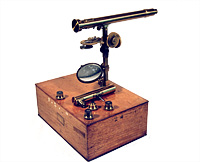
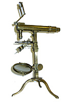
On the same square-section pillar of the microscope which was screwed onto the mahogany case Amici could easily substitute the horizontal tube of the achromatic with the catadioptric tube. Two of these instruments he sent to Paris in 1829 at the price of 1000 francs each: one for the Faculty of Sciences, described by Claude Pouillet in his Elémens de physique expérimentale et de météorologie (download pdf), and the other, pictured here, for the Polytechnic School (photo courtesy of Mr. Rolf Willach). For this latter Amici noted down the following magnification values:
(numbers 1°- 6° indicate the eyepieces):
The third lens with the flat surface towards the object is of short focus, that is 1 ½ internal lines and two scarce external. English Crown. Guinand Flint. Very excellent.
The 3 lenses |
1° |
291 |
With 2 lenses |
1° |
188 |
2° |
518 |
2° |
334 |
||
3° |
758 |
3° |
489 |
||
4° |
1599 |
4° |
1032 |
||
5° |
2629 |
||||
6° |
4299 |
With one |
1° |
84 |
Catadioptric |
1° |
50 |
2° |
149 |
2° |
91 |
||
3° |
218 |
3° |
133 |
||
4° |
283 |
||||
5° |
465 |
 Microscopic objects which accompany microscope n.° 2 for Paris
1. Scales of Papilio Idas. Scopoli Ent. Car.
2. Scales of Papilio Brassicæ
3. Anonymous scales
4. Scales of Papilio Rapæ
5. Scales of Phalæna Pacta
6. Woodworm hairs
7. Mosquito wings
8. Fir wood.
The information sheet for the other Microscope for Paris No. 1 began with this important note:
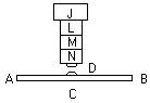
There are three achromatic lenses marked 1.2.3. which can be used together or separately. The combination of all three is the strongest for transparent objects inserted between two plates of glass of the thickness of those provided along with the microscope. The achromatism of this system was calculated taking the thickness of said plates into account between which one commonly encloses small objects like for example infusorians, blood molecules, plant pollens, etc. Without the interposition of one of the two aforementioned subtle plates between the lens and the object, this latter would be magnified all the same but less distinct.
Amici had understood as early as 1827 that there was interference with the cover for the object and he had proposed four solutions to solve this drawback.
 The hemispherical front lens
The first step that led Amici to obtain greater magnification was “the substitution, which he ideated, of a very acute simple lens instead of the third compound lens of the ordinary Series”. It was the hemispherical front lens, first introduced in 1838 in the microscope constructed for Rev. Thomas Romney Robinson, directory of the Armagh Observatory in Ireland. Amici wrote him in May of the same year:
Your microscope is finished [...] I was informed by Sir South that some of the most celebrated Manufacturers of Microscopes in London laughed when they heard that I had asked for high-dispersion Faraday glass. I therefore believed it of benefit to make a combination of achromatised objectives for you with the same glass that I received as a gift a few years ago from Mr. Airy. With this combination, which clearly reveals the tiniest parts of the test objects that I gave to you, you can demonstrate to the artists of the great Capital the value of the highly dispersive glass that the distinguished physicist and their fellow-countryman is capable of constructing.
And on 4 June he wrote to the Astronomer Royal George Biddel Airy
Doctor Robinson, whom I had the pleasure to meet here, has obtained for me a piece of Faraday glass, which I have however not yet received, similar to the one that you graciously sent to me. But I am informed that the most renowned microscope makers in London are laughing, perhaps thinking that with that glass it is not possible to achieve excellent results. To demonstrate just how advantageous is the substance composed by the famous Mr Faraday, I have prepared a five-lens combination with your ancient glass to be sent to Dr. Robinson, who will exhibit it. The incredulous will perhaps be converted.
Another useful document for understanding this important step in the development of the modern achromatic microscope is a letter from Amici to Ottaviano Fabrizio Mossotti, dated 25 October 1855 (cf. A. Meschiari, Corrispondenza di G. B. Amici con Ottaviano Fabrizio Mossotti, “Atti della Fondazione Giorgio Ronchi”, 5-1999):
The object-glasses in my microscopes, consisting of six lenses, three of crown glass and three of Flint glass, glued in pairs with Canada balsam, are constructed with just one quality of crown glass and just one of Flint glass and are achromatic. However, as I said, I found that the series consisting of three pairs of lenses were not the most suitable for obtaining the highest magnifications; in particular because the lower pair facing the object is too large, making it impossible to obtain an extremely small focal length of the system and extremely large aperture. I then thought of replacing the lower pair with a simple lens, namely a half sphere, of any transparent substance, whether crown, flint, balas ruby, diamond, fused rock crystal, etc., and to eliminate the aberrations of this lens by appropriately working the two upper pairs. To do this I procured a Flint glass of extremely high dispersing power, which I was able to obtain from Faraday through the mediation of Airy. The English opticians laughed at this request, but when in 1844 I demonstrated to them in London the superiority of the new construction, they immediately began to imitate it, and likewise the Americans: the French, not caring for it or not understanding the improvement, were left behind.
In his Practical treatise on the use of the microscope John Quekett completes the assertions of Amici:
In the year 1844 Professor Amici visited this country, and brought with him an object-glass of one-seventh of an inch focal length, with an aperture of 112°; this combination was in part composed of Dr. Faraday’s dense glass. Mr. Ross copied Amici’s construction; but found the dense glass so exceedingly soft and fragile, as to render it unfit to receive the high polish essential to the correct performance of any object-glass: he also noticed that Amici’s glasses were much tarnished; he then devised a new construction, whereby, with the ordinary dense glass, he obtained an aperture in the one-eight of 85°, and in the one-twelfth as high as 135°. More recently, Mr. Spencer, of New York, United States, sent to England an object-glass, equivalent to one-twelfth of an inch focal length, and transmitting a pencil of 172°.
The immersion technique
Amici’s most important contribution to the further improvement of the modern achromatic microscope was with the introduction of the immersion technique. He first used water, in 1847, and then different types of oils from the mid-1850s on. He explained this new principle to the micrographer Mr. Mouchet of Rochefort sur Mer in a letter dated 8 January, 1856:
Using the tip of a squirrel-hair paintbrush one has to wet the outside surface of the lower lens in such a way that a small drop of rainwater or better of distilled water remains on it. Another drop of water is placed on the glass plate which covers the microscopic object. Then the two drops are approximated and placed over the viewing point. The object will always appear distinctly defined because the sum of the layer of fluid and of the plate of glass in any case forms a constant and equal thickness. And it is clear that if the fluid had the same refracting power as glass (which is not the case when using water), then the precision of the images of microscopic objects would be mathematically equal, whatever the thickness of the glass plates used.
From 1850 to 1853 the apertures of the series of water-immersion objectives ranged from the 118° of the Panciatichi microscope to the 138° of the Lehmann microscope (1852) to the 140° of the microscope for the Botanical Garden of Modena (1852) to the 160° of the Brachet microscope (1853). Amici wrote about this latter:
The author believes that no other Series previously constructed by opticians is equally powerful as the VI Series consisting of six substances of different refracting and dispersing powers. And considering the distance of about 0.4 between the lower lens and the object (a very large distance in relation to its focal length of 1.74 mm) with an aperture of 160° he believes he has exhibited proof of the superiority of the principle devised by him.
 The first technical sheet that deals with immersion in oil (the type of oil is not specified) dates from 12 January 1856, and therefore refers to a microscope constructed in 1855 (for Rome). The microscope built for Lenoir of Vienna (February 1856) with 18 objectives has a water-immersion V series and a VI series which instead of water uses “clear white, well-prepared poppy-seed oil. If lacking, this oil can be substituted with sweet almond oil” (index of refraction of poppy-seed oil: 1.461). The technical sheet for the Ercolani microscope, which would appear to be earlier than 1855, speaks of immersion in castor oil and glycerine (index of refraction of glycerine: 1.404).
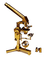
Amici prepared the instrument pictured here, still on display at the History Museum of the University of Pavia, for Prof. Giacomo Sangalli (1821-1897), who taught Anatomy in the Faculty of Medicine and Surgery at that university. In April 1999, this microscope was examined by Dr. Gustavo Merico, who published the results (download pdf).
The microscopes constructed for the Istituto Tecnico in Florence and for Dr. Lamartinière (1859) have a series with immersion in glycerine. Those for Prof. Gabbrielli in Siena (January 1860) and for Prof. Tommasi in Pavia (1860) have a series with immersion in castor oil. The microscope for Prof. Panizza (May 1860) has a series in water and another under aniseed oil (aperture 130°. Index of refraction of aniseed oil: 1.561). There was also a third rock crystal objective lens of smaller aperture used with olive oil (index of refraction of olive oil: 1.461). The microscope for Prof. Tomati in Turin (November 1860) has the series VI under refined walnut 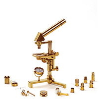 oil, an aperture of 148° and a large focal length. The microscope for Dr. Holdt (April 1862), pictured here, has a maximum aperture of 160° and a series that uses immersion in aniseed and sassafras oil.
Amici wrote about the mixture of aniseed and olive oils: “With these two oils it is possible to compose a fluid with an index of refraction exactly equal to the index of the crown glass that constitutes the lower objective lens of the Series” (index of refraction of the mixture of olive and aniseed oil: 1.496).
Test-objects used by Amici
1826-1829: Papilio Idas, Papilio Brassicae, Papilio Rapae, and Phalena Pact scales, woodworm hairs, mosquito wings, porous tubes of fir wood.
1836-1838: Papilio brassicae, Bombyx Hera, mosquito wings, Noctua Sponsa, Lepisma Sac., Papilio Argus, Papilio Janira, fly muscle fibre.
1845: Noctua sponsa, Papilio janira, Farina fossile.
1847: Male Papilio hermion, Papilio Argus, Tripoli from S. Fiora, Female Papilio janira, Noctua Sponsa. 0.15 mm. and 0.23 mm glass plates.
1850: Test-object sent from Spencer: Grammatophora subtilissima. Rutherfurd sends: Grammatophora Marina and Striatella unipunctata from Cherbourg. Amici sends him soil from Lillhaggsion.
1852: Navicula Amici: his difficult test both dry and under balsam.
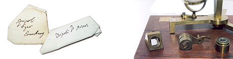
1856: Pleurosigma angulatum in balsam, dry Ceratoneis fasciola, Grammatophora subtilissima, Navicula Amici in balsam, Tripoli from Eger which contains Campylodiscus clypeus and a Navicula, as well as Surirella gemma. Dry: Lycoperdon spores, Hiparchia janira scales, Lycaena scales, Pleurosigma angulatum, Striatella unipunctata Cherbourg. Under balsam: Tripoli from Eger, Grammatophora marina, Tripoli from Santa Fiora, Lapponia (which contains the Navicula Amici).
1857: Navicula Amici, Pleurosigma angulatum. Isthmia, Biddulphia, Campylodiscus, Amphora, Pleurosigma Cocconeis, Navicula, Grammatophora, Ceratoneis fasciola, Eunotia, Surirella gemma.
An undated note written by Amici probably at the beginning of the 1850s contains useful information about the tests Amici used to evaluate the separating power of his microscope objectives:
In the Ceratoneis fasciola, the most difficult English test, whose length is of 0.107 mm, I numbered two hundred striae or transversal lines included from one extremity to the other of the object, thus the distance of each stria from the other is 0.000535mm.
In my most delicate test, that of the fossil infusorians from Lillhaggsion near Umea in Sweden, I counted 250 transversal lines in an individual having a length of 0.073mm, so that the distance between the striae is 0.000292, that is, much smaller than that of the English test.
The striae of my test are the limit of vision of my microscope, and in order to number them I projected the Navicula onto a sheet of lined, thin paper and brought it to a distance where the lines of the paper seemed equally near to each other as those of the Navicula when viewed through the microscope. Then it was easy for me to number the lines included and traced on the piece of paper from one extreme to the other of the projection. For this purpose the lines marked on the backgrounds of some copper engravings can be useful since they are equidistant since traced by a machine.
Over a period of almost half a century, between 1818 and the early 1860s, Amici’s microscopes were used by numerous scientists of international standing in their research work. In the second half of the 1820s in Paris, botanists Adolphe Brongniart and Charles-François Brisseau Mirbel used instruments made by Amici to investigate embryo generation and development in phanerogamic plants. Amici himself had made similar observations using his newly developed reflecting microscope, discovering the pollen tube in 1822 and describing with precision the entire fertilisation process in phanerogamic plants in 1846. The German botanists Hugo von Mohl in Tubingen around the mid-1840s and Wilhelm Hofmeister in Leipzig in the 1850s also used Amici’s lenses to study the formation of the plant embryo.
Also in Paris, Amici’s polarizing microscope was used in the mineralogical and crystallographic investigations of Pierre Armand Dufrénoy, professor of Geology and director of the École des Mines; in the study of the birefringent optical properties of crystals by Alfred Des Cloizeaux, professor of Mineralogy at the École Normale Supérieure; and in the studies of polarisation conducted by Henri de Senarmont, professor of Mineralogy and director of studies at the École des Mines and chief mining engineer from 1848.
In the late 1850s the Bohemian physician Wilhelm Dusan Lambl, the Prague associate of Jan Evangelista Purkyn?, discovered a parasite in the human intestine which he named Cercomonas intestinalis (subsequently Lamblia intestinalis). Josef Hasner, appointed professor of Ophthalmology at the University of Prague in 1856, received an Amici microscope the following year. The Bohemian physician August Breisky, dissector at the Anatomical Institute of Prague, also purchased an Amici microscope in 1857. Lambl reported that physiologist Ernst Wilhlem Brücke of Vienna also used Amici’s microscopes in his laboratory.
In the 1850s, Karl Gotthelf Lehmann, professor of Physiological chemistry in Leipzig and subsequently in Jena, used an Amici microscope with an immersion lens and polarizing apparatus. During the same period in Leipzig the physiologist Otto Funke worked with an Amici microscope. In late 1859, Max Schultze, professor of Anatomy and Histology in Bonn who was working on cell theory, purchased a small Amici microscope for the Bonn Anatomical Institute, of which he was director. This instrument was similar to the one Christian Gottfried Ehrenberg had bought when passing through Florence in October 1858.
In Messina between 1859 and 1860 the young Ernst Haeckel investigated the structure of Radiolaria in plankton using a small Amici microscope mounted on a box and equipped with a water immersion lens.
Pieter Harting, full professor of Pharmacology and plant physiology (from 1846) and one of the leading scholars of the historical development of the microscope, worked at the University of Utrecht with an Amici microscope. After observing and appreciating the instrument owned by Harting, physiologist and ophthalmologist Frans Cornelis Donders also ordered one, which he received in 1849 and used to study pathological anatomy at that university.
Joachim Frederik Schouw, professor of Botany at the University of Copenhagen and a correspondent of Charles Darwin who had an interest in the new science of phytogeography, purchased an Amici achromatic microscope in 1829. In the late 1850s, Jan Van Geuns, professor of Pathology at the School of Medicine in Amsterdam, received an instrument with water and poppy seed oil immersion lenses (160° aperture) and a polarizing apparatus.
British physician Peter Mark Roget, professor of Physiology at the Royal Institution and a member of the Royal Society (he was appointed secretary in 1827 after John Herschel), conducted his research using an Amici reflecting microscope.
In Italy, besides Amici himself, the following used his microscope for their scientific research: Agostino Bassi and Balsamo Crivelli, Luigi Concato, Filippo De Filippi, Giuseppe De Notaris, Giovanni Battista Delponte, Salvadore Gabbrielli, Guglielmo Gasparrini, Carlo Grillenzoni, Pasquale Landi, Carlo Livi, Francesco Magni, Antonio Marcacci, Filippo Pacini, Luigi Paganucci, Bartolomeo Panizza, Giorgio Pellizzari, Luigi Rolando, Giacomo Sangalli, Salvatore Tommasi, Giampaolo Vlacovich, etc.