Instruments
Pocket Microscope
Amici described his new portable or pocket microscope in a Letter to Dr. Francesco Magni (1828-1887), Director of the Florence Anatomy School. This letter was published in the Florence newspaper “Il Tempo”, Giornale di medicina, chirurgia e scienze affini (“Il Tempo”, Journal for Medicine, Surgery and Related Sciences), 3rd issue, March 1858. The instrument was made in two versions, with and without feet.
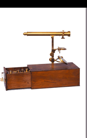
Here is a brief description of the pocket microscope. Figure 1 shows it at normal size. Tube A has the eyepiece at the very top and the objective lens at the very bottom. It slides into another tube B and it can be pushed far enough to give an approximate view of the object (blocked between two slides or placed on top of one only if it is so light in weight that it adheres to the plate) that is placed on the plate CD and blocked with the pressure of the clips E.
The brass plate CD folds back onto itself at D and has not only the tube B attached to it, but also a micrometer screw button F. By rotating this latter right or left the plate CD is moved towards and away from the objective, so that with delicacy the object can be brought to the right point to be seen distinctly.
Illumination is obtained using a triangular glass prism G which has one of the smaller sides of a spherical form. Situated in the middle of a ring attached to a cylindrical stem which is inserted into the flexible tube H, it can be placed more or less near the object, varying the intensity of shine. And since it turns around its stem as well, the illumination can be central or oblique, as needed. It is clear that tube B is inclined as it is pictured here if light is to be taken from a window or from a candle above the instrument, and the best direction can easily be found. But since one of the user’s hands is occupied in holding the microscope, he only has the other free to move the object, which actually is not an obstacle to observations for anyone that has experience and dexterity in carrying them out.
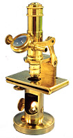
However, should one wish to have both hands free, a different apparatus, like the one in Figure 2, would be needed.
Here the tube which contains the lenses is vertical and sustained by a metal arm M. Plate CD is turned over so that the plane on which the slides containing the objects sits and the clips E which block them are on top now. The micrometric screw F moves to under. Under there is also a pillar or tube blocked by screws and open on one side to allow the light to enter. The light is concentrated and reflected vertically by the mirror S and illuminates the item that is being studied. The microscope is completed by a round base of a certain weight.
The mechanical disposition of the two small instruments which I have described seems to have achieved the aim that I had, that was to construct them with the greatest possible economy but without limiting too much their convenience and effectiveness. The forms can certainly be varied in many ways yet, but I have only created what I considered to be most useful to the average observer.
The optical parts of them, as I mentioned, are composed of an eyepiece and an object-glass. The eyepiece is positive and of the Ramsden type even if not identical to it. Mine has one of its two lenses composed of two glasses of different dispersion, and I demonstrated it to the first Congress of Italian Scientists held in Pisa. It provides a wider and more achromatic field of vision from the common eyepieces. The area visible in the pocket microscope is about four times the area which the ordinary eyepieces used by producers provide. The same advantage of a wide field can be obtained with a negative eyepiece. There is no need to hide the fact that in case of too much illumination the objects located near the border of the large field will not be as sharp as those which are located towards the centre, but it is always useful to be able to take in all the objects in a single glance in
order to be aware of the existence of exactly what one is looking for and wanting to see with greater distinction. That object can then be moved into the centre of the field.
The object-glass is composed of two lenses, each of which made of two qualities of glass and combined together. Its focal distance allows one to look even under glass as thick as from two to three millimetres with a linear magnification of two hundred times at lest, and the majority of the test objects cited by the micrographers can be seen. For example, the longitudinal lines in the scales of the wings of P. Brassica, P. Argus, etc.The interrupted lines in the scales of the Podura plumbea; the striae of the Navicula Hippocampus, which can be considered the limit of the penetrating power of this objective, can all be seen quite well. It is no excess to point out that none of these minute particulars could be seen with the microscopes constructed last century or at the beginning of this by the famous foreign opticians; microscopes like the ones which the Physics Museum has, and which I myself have, nonetheless served the renowned scientists Felice Fontana and Bonaventura Corti in making many important observations. It would therefore be hoped that a portable microscope, as reduced as possible in volume but with sufficient optical force, accessible even to those with limited funds and made popular would prove to be useful especially in medicine and the natural sciences.
Note. The weight of the microscope indicated in Figure 1 is less than five Florentine ounces = about 14 decagrams. It costs 50 francs. The microscope pictured in Figure 2 costs 60 francs.
 The microscope pictured here is kept in the Department of Anatomy and Histology of the University of Modena. It is 12.30cm high and it has a plate for the objects measuring 6.30 x 3.70 cm. It has an eyepiece with two lenses and a 1,2,3 compound object-glass. Giuseppe Favaro mentions it in the article Il microscopio di G. B. Amici dell’Istituto Anatomico di Modena, “Rassegna per la Storia della Università di Modena e della cultura superiore modenese”, I-1929 (The microscope of G.B. Amici in the Anatomical Institute of Modena, “Review of the history of the University of Modena and of higher education in Modena”).